
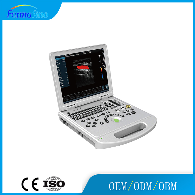
4D Color Doppler Ultrasound Therapy Machines
Original price was: $5,999.00.$4,999.00Current price is: $4,999.00. & Free Shipping
Free shipping on orders over $50!
- Satisfaction Guaranteed
- No Hassle Refunds
- Secure Payments
Description
Essential details
Place of Origin:
China
Brand Name:
FarmaSino/FarmaMed/BodTrust/OEM
Model Number:
FDW-L5
Power Source:
Electric
Warranty:
3 years
After-sale Service:
Online technical support
Material:
Plastic, Steel
Shelf Life:
2 years
Quality Certification:
CE ISO
Instrument classification:
Class II
Safety standard:
GB 9706.1
Product name:
FDW-L5 Color Doppler Ultrasound Machine
Display:
15.6 Inch HD LED Screen
Keyword:
Portable Color Ultrasound
Battery:
Built-in large capacity lithium battery (detachable)
Storage:
120G Memory
USB ports:
2
OS:
Windows
Supply Ability
Supply Ability500 Set/Sets per Year
Packaging & delivery
Packaging DetailsPortable ultrasound packed with cartonPortShanghai/QingdaoLead time:
| Quantity(sets) | 1 – 5 | >5 |
| Est. time (days) | 10 | To be negotiated |
Color Doppler Ultrasound Machine
Products Description
High resolution medical 15.6 inch display.
Spectrum envelope function.
One-click automatic optimization.
Rich user-defined functions.
Built-in large capacity lithium battery (detachable).
Product Description
Micron Imaging Technology
Micron imaging technology, real-time tracking of specific signals at the edges of different tissues, to achieve edge enhancement,
and monitor each pixel at the same time; optimize the internal signal of the organization and perfectly integrate the edge
information and the internal pixel information of the organization to restore the real and delicate, excellent level contrast
Two-dimensional image.
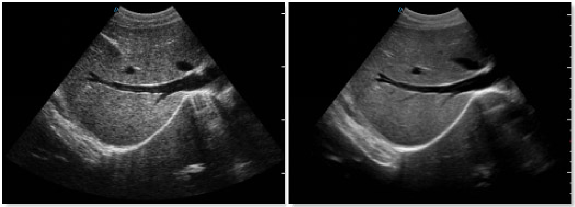
Harmonic Imaging Technology(THI)
It improves image clarity by improving tissue contrast resolution, spatial resolution, and eliminating near-field artifacts.
It is mainly used for the diagnosis of cardiovascular and abdominal diseases. It plays an important role in evaluating the lesion area and boundary division of patients with imaging difficulties. The technology has been fully approved by clinicians.
Harmonic technology retains the second harmonic signal to the greatest extent on the basis of removing the fundamental signal, which increases the signal strength by more than 30% compared with the traditional signal processing, reduces noise and artifacts, and improves the contrast resolution of
tissue images.
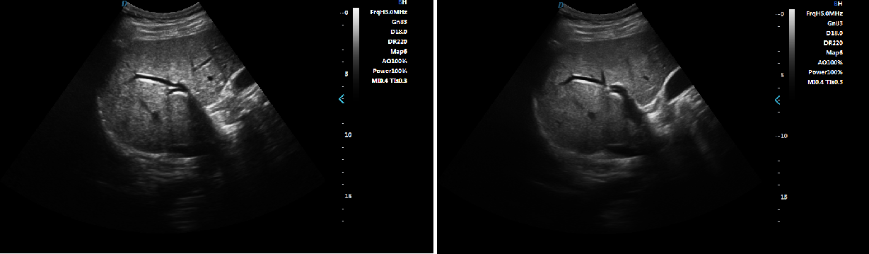
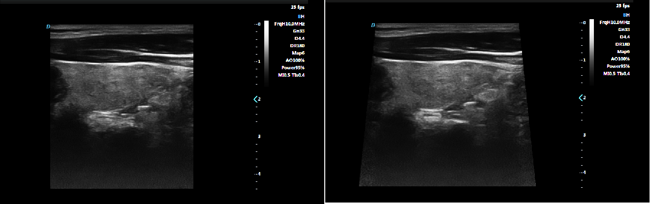
Trapezoid Imaging
Trapezoid imaging is a kind of expanded imaging, which is transformed into a trapezoid on the basis of the original rectangle, and
the left and right sides are expanded to a certain extent, achieving a wider field of view. The principle of ultrasound imaging is
to scan the human body with ultrasonic sound beams, and obtain images of internal organs by receiving and processing the reflected
signals.
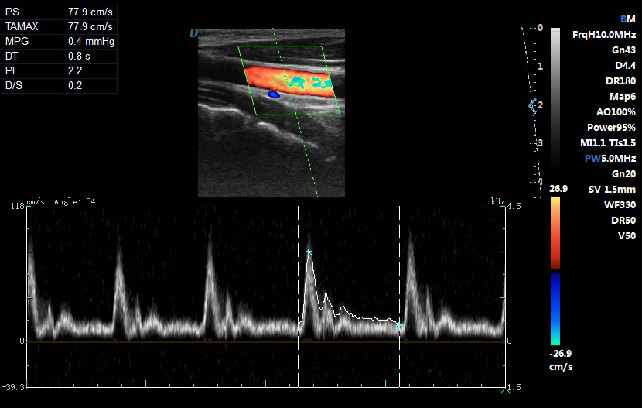
Automatic Spectrum Tracking Measurement Technology
Ultrasound Doppler technology is used in the ultrasound system for examining the heart and arteries and veins. It is necessary to extract relevant parameters from the Doppler spectrogram to evaluate the hemodynamic status of the heart and blood vessels. The disadvantage of manual detection is that the operator’s marking of the peak velocity is
relatively monotonous and time-consuming, with poor repeatability and low estimation accuracy; and during the detection, in order to mark the peak velocity, the operator needs to interrupt the acquisition of Doppler signals, which makes it impossible to estimate in real time. This host contains an automatic envelope detection module, which can automatically track the time-related changes of the peak blood flow velocity and average velocity, and display them in real time on the Doppler spectrogram.
| 1 | 7.5Mhz(2.0-10.0Mhz) linear probe |
| 2 | 7.0MHz(2.0-10.0Mhz) trans-vaginal probe |
| 3 | 2.0MHz(2.0-10.0Mhz) Microconvex probe |
and monitor each pixel at the same time; optimize the internal signal of the organization and perfectly integrate the edge
information and the internal pixel information of the organization to restore the real and delicate, excellent level contrast
Two-dimensional image.
tissue images.
the left and right sides are expanded to a certain extent, achieving a wider field of view. The principle of ultrasound imaging is
to scan the human body with ultrasonic sound beams, and obtain images of internal organs by receiving and processing the reflected
signals.
relatively monotonous and time-consuming, with poor repeatability and low estimation accuracy; and during the detection, in order to mark the peak velocity, the operator needs to interrupt the acquisition of Doppler signals, which makes it impossible to estimate in real time. This host contains an automatic envelope detection module, which can automatically track the time-related changes of the peak blood flow velocity and average velocity, and display them in real time on the Doppler spectrogram.
|
1
|
7.5Mhz(2.0-10.0Mhz) linear probe
|
|
2
|
7.0MHz(2.0-10.0Mhz) trans-vaginal probe
|
|
3
|
2.0MHz(2.0-10.0Mhz) Microconvex probe
|

Related products
-
-
Sale!

Comfortable Nursing Bed Manual Adjustable Hospital Bed With Mattress
Original price was: $200.00.$188.00Current price is: $188.00.
Reviews
There are no reviews yet.